
X-Ray Microscope & Micro/Nano-Computed Tomography
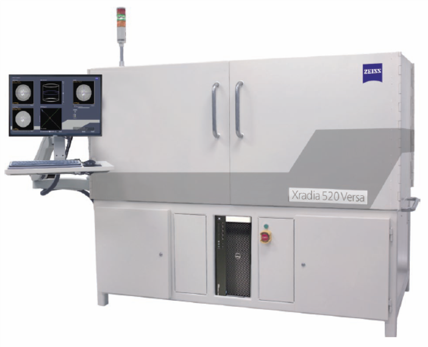
Zeiss Xradia 520 Versa X-ray Microscope (XRM) and Nano/Micro-Computed Tomography System
Why XRM?
XRM has emerged as the gold standard in materials imaging. XRM provides a unique, non-destructive means to image and differentiate internal material micro- and nanostructures using unmatched resolution and phase contrast. Internal structure plays a critical role in physical and mechanical behavior of both natural and engineered materials, enabling the design of complex synthetics and biomimetics.
How does it work?
XRMs create cross-sections of scanned objects using X-rays that can be combined to generate a 3D virtual model. Non-destructive 3D XRMs enable characterization of time-dependent microstructural development and the evolution of properties over time, 4D.
Why the ZEISS Xradia XRM?
A unique paring of X-ray source and objective turret enables the ZEISS XRM to achieve unparalleled resolution (down to 700 nm) and phase contrast in both small and large (300 mm) samples. New advances in XRM now permit in situ imaging of material behavior in 3D/4D under controlled temperature, compression, and tension, enabling previously unobservable deformation and failure. Beyond quantifying physical and mechanical properties in situ, image data can be directly imported into numerical simulations and manipulated with stress, strain, temperature, pressure, and fluid flow to computationally model and observe microscale behaviors.
Technical Details.
The ZEISS Xradia 520 Versa system includes a 160 kV high-energy microfocus X-ray source and staging platform, a 2kx2k high-resolution 16-bit CCD digital camera assembly, a ZEISS 0.4X large field of view (FOV) camera assembly, a ZEISS high contrast, low resolution 4X detector, a ZEISS high contrast, ultra-high resolution 20X detector for high energy and 4-axis tomography, a motorized precision sample stage with a load capacity of 15 kg and 360° capability, automatic filter changer with 12 standard filters, a radiation-safe four-door steel enclosure with redundant safety interlock, temperature control, and “X-ray On” indicator light, and interior camera. The system comes with alignment and calibration standards for quality control. The sample chamber can accomodate samples from < 1 mm and up to 30 cm in diameter. Samples can be imaged while contained in X-ray transparent containment to permit evaluation even while submerged in a fluid.
In Situ Interface Kit.
An In Situ Interface Kit for the Xradia 520 Versa is complete with a CT5000-RT in situ tension-compression stage with a

Computer Workstations and Software.
Two dedicated computer workstations are avaialble for XRM instrument operations and post-processing and 3D image reconstruction. An advanced imaging software package, Dragonfly Pro, provides a common workspace for multi-scale (nm to cm) correlative microscopy with a simple user interface and is fully customizable with Python.